Phone: 085912 91896 Email: info@spinespecialist.co.in

Spinal Stenosis Treatment in Mumbai
Expert Diagnosis and Personalised Treatment for Spinal Stenosis by Spine Surgeon Dr Satyen Mehta
Spine Surgeon | 25+ Years of Experience | Consultation Locations: Chembur | Khar | Mulund
Spinal stenosis is a common age-related condition in which the spaces within the spine become narrower, placing pressure on the spinal cord or spinal nerve roots. This narrowing can lead to persistent back or neck pain, numbness, tingling, leg pain, weakness, balance problems, and difficulty walking. While symptoms often develop gradually, they may progressively interfere with everyday activities if left untreated.
The condition most commonly affects the lumbar spine (lower back) and the cervical spine (neck). Some patients experience mild symptoms that improve with physiotherapy, activity modification and medication, while others develop significant nerve compression requiring advanced treatment.
Dr Satyen Mehta is a dedicated spine surgeon in Mumbai with over 25 years of experience in diagnosing and treating spinal stenosis and other complex spine disorders. Every patient undergoes a detailed clinical evaluation and careful review of imaging before treatment is recommended. Depending on the severity of the condition, treatment may include medication, physiotherapy, image-guided spinal injections, minimally invasive spine surgery or endoscopic spine surgery when clinically indicated.
What is Spinal Stenosis?
Spinal stenosis is a condition in which the spinal canal narrows, reducing the space available for the spinal cord and spinal nerves. As this space becomes smaller, the nerves may become compressed, leading to pain, numbness, weakness or difficulty walking.
The spine is made up of a series of vertebrae stacked one above another. Running through the centre of these vertebrae is the spinal canal, which protects the spinal cord and the nerves travelling to different parts of the body.
With ageing or certain spinal disorders, structures around the spinal canal gradually enlarge or thicken. These changes reduce the available space for the nerves and may eventually produce symptoms of spinal stenosis.
Although spinal stenosis can occur anywhere along the spine, it most frequently affects:
- Lumbar spine (lower back) – causing lower back pain, leg pain and walking difficulty.
- Cervical spine (neck) – causing neck pain, arm symptoms, hand weakness and balance problems.
- Thoracic spine (mid-back) – uncommon but may affect walking and lower limb function.
The severity of symptoms depends on the degree of nerve compression rather than the amount of narrowing seen on imaging alone. This is why treatment decisions are always based on the patient’s symptoms, neurological examination and MRI findings together.
Understanding Your Spine
To understand spinal stenosis, it helps to know how the spine is structured and how the spinal nerves travel through it.
The spine has three important functions:
- Supports the weight of the body.
- Protects the spinal cord and spinal nerves.
- Allows controlled movement while maintaining stability.
It is made up of vertebrae separated by intervertebral discs, which act as shock absorbers during daily activities. Small joints called facet joints guide spinal movement, while strong ligaments provide additional stability.
Running through the centre of the vertebrae is the spinal canal, a protective tunnel that contains the spinal cord in the neck and upper back, and the nerve roots in the lower back.
Healthy spinal structures allow these nerves to move freely. However, when the canal becomes narrow because of age-related degeneration, thickened ligaments, enlarged joints or disc bulges, pressure develops on the nerves, leading to the symptoms of spinal stenosis.
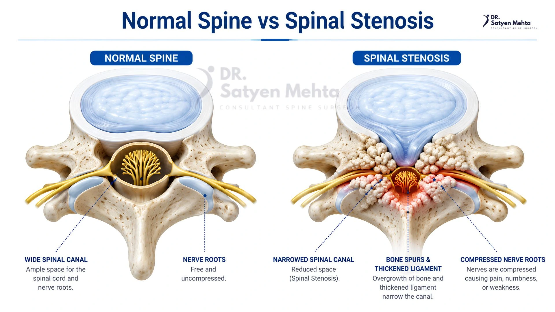
Normal Spine vs Spinal Stenosis
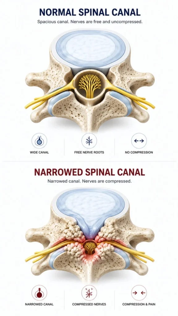
In a healthy spine, the spinal canal provides sufficient space for the spinal cord and spinal nerves to pass without restriction.
In spinal stenosis, this space gradually decreases due to structural changes such as:
- Bulging or degenerated intervertebral discs
- Enlargement of the facet joints due to arthritis
- Thickening of the ligamentum flavum
- Formation of bone spurs (osteophytes)
- Vertebral slippage (spondylolisthesis)
As the available space becomes smaller, the nerves become compressed, resulting in inflammation and reduced nerve function.
This explains why many patients experience symptoms that become worse during standing or walking but improve after sitting or bending forward. Flexing the spine temporarily increases the available space around the nerves, providing temporary relief.
Types of Spinal Stenosis
Spinal stenosis is classified according to the region of the spine where narrowing occurs.
Lumbar Spinal Stenosis
Lumbar spinal stenosis affects the lower back and is the most common type of spinal stenosis.
Patients often experience:
- Lower back pain
- Pain radiating into one or both legs
- Numbness or tingling in the feet
- Weakness while walking
- Difficulty standing for prolonged periods
- Relief after sitting or bending forward
Walking limitation caused by lumbar spinal stenosis is often described as neurogenic claudication, a characteristic symptom that helps distinguish it from other causes of leg pain.
Cervical Spinal Stenosis
Cervical spinal stenosis occurs when the spinal canal narrows in the neck.
As the spinal cord becomes compressed, patients may experience:
- Neck pain
- Pain radiating into the shoulders or arms
- Tingling or numbness in the hands
- Reduced grip strength
- Difficulty with fine hand movements
- Balance problems while walking
In advanced cases, cervical spinal stenosis may lead to cervical myelopathy, a condition requiring prompt evaluation to prevent permanent neurological damage.
Thoracic Spinal Stenosis
Thoracic spinal stenosis is relatively uncommon because the mid-back is naturally more stable than the neck or lower back.
When it occurs, symptoms may include:
- Mid-back pain
- Weakness in the legs
- Difficulty walking
- Balance problems
- Changes in bowel or bladder function in severe cases
Because thoracic stenosis can affect the spinal cord directly, it requires careful evaluation and timely management.
Why Does the Spinal Canal Become Narrow?
Spinal stenosis usually develops gradually over many years. Rather than being caused by a single event, it often results from a combination of age-related changes affecting the spine.
Common mechanisms include:
Disc Degeneration
As spinal discs lose water content with age, they become thinner and less effective as shock absorbers. Reduced disc height alters spinal mechanics and contributes to narrowing around the nerves.
Bone Spurs (Osteophytes)
Arthritic changes stimulate the formation of extra bone around the vertebrae and facet joints. These bone spurs may project into the spinal canal and compress nearby nerves.
Thickening of the Ligaments
The ligamentum flavum, which helps stabilise the spine, may gradually become thickened and less flexible. This further reduces the space available within the spinal canal.
Facet Joint Arthritis
Degeneration of the facet joints can enlarge these joints and contribute to narrowing of both the spinal canal and the openings through which spinal nerves exit.
Spondylolisthesis
When one vertebra slips forward over another, the alignment of the spine changes, reducing the available space for the nerves and increasing the likelihood of spinal stenosis.
Congenital Narrow Spinal Canal
Some individuals are born with a naturally smaller spinal canal. Even mild age-related changes may then produce symptoms earlier than expected.
Key Takeaway
Spinal stenosis is rarely caused by a single abnormality. In most patients, it results from several age-related changes occurring together. This is why successful treatment begins with identifying which structures are compressing the nerves and determining whether those changes correspond with the patient’s symptoms and clinical examination.
What Causes Spinal Stenosis?
Spinal stenosis most commonly develops as part of the natural ageing process. Over time, the structures that support the spine undergo gradual wear and tear, reducing the available space for the spinal cord and spinal nerves. In many patients, more than one structural change contributes to nerve compression.
Understanding the underlying cause is important because treatment is directed at relieving the source of compression rather than simply controlling pain.
Age-Related Degeneration
The most common cause of spinal stenosis is age-related degeneration of the spine. As the intervertebral discs lose water content and height, the mechanics of the spine change. This places additional stress on the facet joints and supporting ligaments, gradually narrowing the spinal canal.
Disc Bulge and Herniated Disc
A bulging or herniated disc may protrude into the spinal canal or neural foramen, reducing the space available for the spinal nerves.
In younger adults, a slipped disc is often the primary cause of nerve compression, whereas in older adults it frequently occurs alongside other degenerative changes.
Bone Spurs (Osteophytes)
As arthritis develops in the spine, the body attempts to stabilise the affected joints by producing extra bone known as osteophytes or bone spurs.
These bone spurs may project into the spinal canal or nerve exit pathways, compressing nearby nerves.
Thickening of the Ligamentum Flavum
The ligamentum flavum is a strong ligament lining the back of the spinal canal.
With age, it may become thicker and less elastic, further narrowing the canal and increasing pressure on the spinal nerves.
Facet Joint Arthritis
Facet joints guide movement between adjacent vertebrae. Degeneration of these joints can lead to enlargement, inflammation and additional narrowing around the nerves.
Facet arthritis commonly contributes to lumbar spinal stenosis in older adults.
Spondylolisthesis
When one vertebra slips forward over another, the alignment of the spine changes. This instability can narrow both the spinal canal and the openings through which the nerves leave the spine.
Previous Spine Injury or Surgery
Fractures, spinal trauma or previous spinal surgery may alter normal spinal anatomy and occasionally contribute to spinal stenosis.
Congenital Spinal Stenosis
Some individuals are born with a naturally narrow spinal canal. They may remain symptom-free for many years until age-related degeneration further reduces the available space around the nerves.
Risk Factors for Spinal Stenosis
Although ageing is the strongest risk factor, several other factors increase the likelihood of developing spinal stenosis.
These include:
- Age over 50 years
- Osteoarthritis of the spine
- Previous spinal injury
- Congenital narrow spinal canal
- Repetitive heavy lifting
- Occupations involving prolonged standing
- Sedentary lifestyle
- Obesity
- Smoking
- Osteoporosis
- Previous spinal surgery
While some risk factors cannot be changed, maintaining a healthy weight, remaining physically active and improving posture may help reduce stress on the spine.
Symptoms of Spinal Stenosis
Symptoms vary depending on the location and severity of nerve compression. Some patients experience only occasional discomfort, while others develop progressive neurological symptoms that interfere with daily life.
Symptoms of Lumbar Spinal Stenosis
Lumbar spinal stenosis affects the lower back and is the most common type of spinal stenosis.
Patients may experience:
- Lower back pain
- Pain radiating into one or both legs
- Pain while walking
- Numbness in the legs or feet
- Tingling or burning sensation
- Leg weakness
- Difficulty standing for long periods
- Reduced walking distance
- Relief after sitting or bending forward
One of the most characteristic symptoms is neurogenic claudication, where walking gradually becomes painful but symptoms improve after sitting or leaning forward.
Unlike muscle pain, the discomfort often returns after walking a similar distance again.
Symptoms of Cervical Spinal Stenosis
When narrowing occurs in the neck, pressure may develop on both the spinal cord and the spinal nerves.
Common symptoms include:
- Persistent neck pain
- Pain radiating into the shoulder or arm
- Tingling or numbness in the hands
- Reduced grip strength
- Difficulty buttoning clothes or writing
- Hand clumsiness
- Balance problems while walking
- Frequent falls
- Weakness in the arms or legs
Progressive spinal cord compression may result in cervical myelopathy, which requires prompt specialist evaluation.
Symptoms That Require Immediate Medical Attention
Although uncommon, certain symptoms may indicate severe nerve or spinal cord compression.
Seek urgent medical evaluation if you develop:
- Progressive weakness in the legs
- Difficulty walking that rapidly worsens
- Loss of bladder control
- Loss of bowel control
- Numbness around the groin (saddle anaesthesia)
- Sudden loss of balance
- Rapid deterioration in hand function
These symptoms require immediate assessment because delayed treatment may result in permanent neurological damage.
Spinal Stenosis vs Sciatica
Many patients use the terms spinal stenosis and sciatica interchangeably, but they are not the same condition.
Sciatica is a symptom, whereas spinal stenosis is one of the conditions that can cause it.
Symptoms of Spinal Stenosis
Many patients use the terms spinal stenosis and sciatica interchangeably, but they are not the same condition.
Sciatica is a symptom, whereas spinal stenosis is one of the conditions that can cause it.
Spinal Stenosis
Narrowing of the spinal canal or nerve passage
A structural spinal condition
Often affects both legs in lumbar stenosis
Often affects both legs in lumbar stenosis
Commonly develops gradually with age
May cause walking limitation
Sciatica
Pain caused by irritation or compression of the sciatic nerve
A symptom rather than a diagnosis
Usually affects one leg
Commonly develops gradually with age
Often begins suddenly, especially with a slipped disc
Typically causes shooting pain down the leg
When Should You Consult a Spine Surgeon?
Many patients continue taking painkillers for months without understanding why their symptoms persist.
A specialist spine evaluation should be considered if:
- Back or neck pain lasts longer than six weeks
- Pain repeatedly returns despite treatment
- Walking distance gradually decreases
- Pain begins travelling into the arm or leg
- Numbness or tingling develops
- Weakness affects daily activities
- Balance becomes impaired
- Sleep is disturbed because of pain
- Physiotherapy and medication fail to provide lasting relief
Early evaluation allows the underlying cause to be identified before prolonged nerve compression results in permanent neurological damage.
How Dr Satyen Mehta Evaluates Patients with Spinal Stenosis
Not every patient with spinal stenosis requires surgery, and not every MRI showing spinal narrowing explains a patient’s symptoms.
For this reason, Dr Satyen Mehta follows a structured evaluation before recommending treatment.
The assessment includes:
- A detailed discussion of symptoms and their progression
- Review of previous treatments and response
- Clinical examination
- Neurological assessment
- Assessment of walking pattern and balance
- Evaluation of muscle strength and reflexes
- Correlation of examination findings with MRI or other imaging
Only after combining these findings is an individualised treatment plan developed.
This careful approach helps distinguish patients who are likely to improve with conservative treatment from those who may benefit from advanced interventions such as minimally invasive or endoscopic spine surgery.
How is Spinal Stenosis Diagnosed?
An accurate diagnosis is the foundation of successful treatment. While MRI scans are valuable, spinal stenosis cannot be diagnosed based on imaging alone. Many people have age-related narrowing of the spinal canal without experiencing symptoms, while others develop significant pain despite relatively modest changes on MRI.
For this reason, Dr Satyen Mehta combines a detailed medical history, clinical examination and imaging findings before recommending any treatment.
Medical History
The consultation begins with understanding how the symptoms developed and how they affect daily life.
Important questions include:
- When did the pain begin?
- Does the pain travel into one or both legs?
- How far can you walk before the pain starts?
- Does sitting or bending forward provide relief?
- Is there numbness or tingling?
- Have you noticed weakness in your legs?
- Does the pain disturb your sleep?
- Have medicines or physiotherapy helped?
- Have you undergone previous spine treatment or surgery?
The answers often provide valuable clues about the level and severity of nerve compression.
Clinical Examination
A detailed physical examination helps identify the affected spinal level and assess nerve function.
During the examination, Dr Satyen Mehta evaluates:
- Posture and spinal alignment
- Walking pattern (gait)
- Balance and coordination
- Range of spinal movement
- Muscle strength
- Sensation in the upper and lower limbs
- Tendon reflexes
- Signs of spinal cord compression
- Nerve tension signs where appropriate
In many cases, the examination findings help explain the patient’s symptoms even before imaging is reviewed.
MRI Scan
MRI is the investigation of choice for spinal stenosis because it provides detailed images of the spinal canal, discs, nerves and surrounding soft tissues.
An MRI can identify:
- Narrowing of the spinal canal
- Compression of spinal nerves
- Disc bulges or herniation
- Thickened ligaments
- Facet joint enlargement
- Degenerative disc disease
- Spondylolisthesis
- Spinal cord compression
MRI also helps determine the exact spinal level responsible for the patient’s symptoms and assists in planning treatment when surgery becomes necessary.
X-Rays
Although X-rays do not show the spinal cord or nerves, they remain useful for assessing the bony alignment of the spine.
Standing X-rays help identify:
- Degenerative changes
- Vertebral alignment
- Spondylolisthesis
- Scoliosis
- Fractures
- Spinal instability
Dynamic flexion-extension X-rays may be recommended when instability is suspected.
CT Scan
CT scans provide detailed images of the bony structures of the spine and may be recommended when MRI cannot be performed or when additional information is required for surgical planning.
Electromyography (EMG) and Nerve Conduction Studies
In selected patients, nerve conduction studies and electromyography (EMG) may help distinguish spinal nerve compression from other neurological disorders such as peripheral neuropathy or nerve entrapment.
These tests are not required for every patient but may be useful when the diagnosis remains uncertain.
MRI Findings Must Match Your Symptoms
One of the biggest misconceptions about spinal stenosis is that an MRI automatically determines whether surgery is required.
This is not true.
Many adults over the age of 50 have MRI findings such as disc degeneration, disc bulges or spinal canal narrowing without experiencing significant symptoms. Similarly, some patients with relatively mild MRI changes may develop severe pain or neurological weakness.
For this reason, treatment decisions are never based on MRI findings alone.
Dr Satyen Mehta correlates three equally important factors before recommending treatment:
- Your symptoms
- Clinical examination findings
- MRI or other imaging studies
Only when these findings match is a treatment plan developed.
This approach helps avoid unnecessary surgery while ensuring that patients with significant nerve compression receive timely treatment.
Conditions That Can Mimic Spinal Stenosis
Not every patient with leg pain or walking difficulty has spinal stenosis.
Several other conditions may produce similar symptoms and should be considered before treatment begins.
These include:
Slip Disc (Lumbar Disc Herniation)
A slipped disc can compress a lumbar nerve root and cause pain radiating into the leg, closely resembling spinal stenosis.
Sciatica
Sciatica is a symptom caused by irritation of the sciatic nerve. It may occur due to spinal stenosis, a slipped disc or other conditions affecting the lumbar spine.
Peripheral Neuropathy
Diabetes and certain neurological disorders may cause numbness, burning sensations and weakness in the feet without any spinal compression.
Hip Arthritis
Pain arising from the hip joint may radiate into the thigh or groin and occasionally resemble symptoms originating from the spine.
Vascular Claudication
Reduced blood supply to the legs due to peripheral arterial disease can also cause pain while walking.
Unlike spinal stenosis, the symptoms usually do not improve by bending forward and are related to reduced circulation rather than nerve compression.
Recognising these differences is important because the treatment for each condition is entirely different.
Can Spinal Stenosis Be Treated Without Surgery?
Yes.
Many patients with spinal stenosis improve without surgery, particularly when symptoms are mild to moderate and neurological function remains stable.
The goals of non-surgical treatment are to:
- Reduce pain and inflammation
- Improve mobility
- Strengthen the muscles supporting the spine
- Maintain independence in daily activities
- Delay or avoid surgery whenever appropriate
Treatment is individualised according to the severity of symptoms, MRI findings and the patient’s overall health.
Non-Surgical Treatment for Spinal Stenosis
Activity Modification
Patients are encouraged to remain active while avoiding activities that significantly worsen symptoms.
Simple modifications such as taking regular walking breaks, avoiding prolonged standing and maintaining proper posture may help reduce nerve irritation.
Medications
Medication may be prescribed to reduce inflammation and relieve pain during the initial stages of treatment.
Depending on the patient’s symptoms, treatment may include:
- Pain-relieving medication
- Anti-inflammatory medication
- Medication for nerve-related pain
- Muscle relaxants where appropriate
Medication should always be taken under medical supervision.
Physiotherapy
Physiotherapy plays an important role in improving flexibility, muscle strength and spinal stability.
A rehabilitation programme may include:
- Core strengthening exercises
- Flexibility exercises
- Balance training
- Walking rehabilitation
- Postural correction
- Stretching exercises
- Functional strengthening
The programme is tailored to the patient’s symptoms and physical ability.
Lifestyle Modification
Long-term management focuses on reducing stress on the spine and preventing further degeneration.
Patients are advised to:
- Maintain a healthy body weight
- Stay physically active
- Stop smoking
- Improve workplace ergonomics
- Avoid prolonged sitting
- Use safe lifting techniques
These changes support both symptom relief and long-term spinal health.
Image-Guided Spine Injections
Some patients continue to experience significant pain despite medication and physiotherapy.
In selected cases, image-guided spinal injections may help reduce inflammation around the compressed nerves and provide temporary symptom relief.
These procedures are performed under imaging guidance for improved precision.
Common options include:
Epidural Steroid Injection
A corticosteroid is injected around the affected nerve roots to reduce inflammation and improve pain.
This may help patients participate more effectively in physiotherapy and postpone surgery in appropriate cases.
Selective Nerve Root Block
A targeted injection around a specific nerve root may be used when symptoms arise predominantly from a single compressed nerve.
It may also assist in confirming the source of pain before surgery.
Facet Joint Injection
Patients with significant facet joint arthritis contributing to back pain may benefit from image-guided facet joint injections as part of a comprehensive treatment plan.
Radiofrequency Ablation
In selected patients with chronic pain arising from the facet joints, radiofrequency ablation may provide longer-lasting pain relief by interrupting pain signals from the affected nerves.
It is important to understand that these procedures relieve inflammation and pain but do not reverse the structural narrowing responsible for spinal stenosis.
They are recommended only after careful clinical assessment and are often combined with physiotherapy and lifestyle modification.
Key Takeaway
The majority of patients with spinal stenosis do not require immediate surgery.
A structured programme combining clinical assessment, physiotherapy, medication, lifestyle modification and, when appropriate, image-guided injections can successfully relieve symptoms in many patients.
Surgery is considered only when these measures fail to provide adequate relief or when progressive nerve compression begins to affect muscle strength, walking ability or spinal cord function.
When is Surgery Required for Spinal Stenosis?
One of the most common questions patients ask is, “Do I need surgery?”
The answer depends on the severity of nerve compression, the impact on daily activities and whether non-surgical treatment has provided adequate relief.
Most patients with spinal stenosis do not require immediate surgery. Many experience significant improvement with medication, physiotherapy, activity modification and image-guided spinal injections.
However, surgery may be recommended when symptoms continue to progress despite appropriate conservative treatment or when nerve compression begins to affect neurological function.
Surgery may be considered if you have:
- Persistent leg pain despite adequate non-surgical treatment
- Difficulty walking because of neurogenic claudication
- Progressive numbness or muscle weakness
- Reduced walking distance affecting quality of life
- Loss of balance due to spinal cord compression
- Symptoms of cervical myelopathy
- Bladder or bowel dysfunction caused by severe nerve compression
- MRI findings that correlate with your clinical symptoms
The primary objective of surgery is to relieve pressure on the compressed nerves, reduce pain, improve walking ability and prevent further neurological deterioration.
Surgical Treatment Options for Spinal Stenosis
The choice of surgery depends on the location of stenosis, the number of affected spinal levels, spinal stability and the patient’s overall health.
Rather than performing the same operation for every patient, Dr Satyen Mehta recommends the procedure that offers the safest and most effective outcome based on the individual’s condition.
Endoscopic Spine Surgery
For carefully selected patients, Endoscopic Spine Surgery allows nerve decompression through a very small skin incision using specialised instruments and a high-definition endoscopic camera.
Potential advantages include:
- Small skin incision
- Minimal muscle damage
- Reduced blood loss
- Less post-operative pain
- Shorter hospital stay
- Faster recovery
- Earlier return to daily activities
However, endoscopic surgery is not suitable for every patient. The decision depends on the underlying spinal pathology and MRI findings.
Minimally Invasive Spine Surgery (MISS)
Minimally invasive spine surgery uses specialised instruments and modern surgical techniques to minimise disruption of normal muscles and soft tissues while effectively decompressing the nerves.
Compared with traditional open surgery, suitable patients may benefit from:
- Smaller incisions
- Reduced tissue trauma
- Less blood loss
- Faster mobilisation
- Shorter hospital stay
- Earlier rehabilitation
The choice between minimally invasive and conventional surgery depends on the patient’s diagnosis and surgical goals.
Decompression Surgery
The goal of decompression surgery is to create more space for the compressed nerves by removing the structures responsible for narrowing the spinal canal.
Depending on the patient’s condition, decompression may involve:
- Laminectomy
- Laminotomy
- Foraminotomy
- Removal of hypertrophied ligamentum flavum
- Removal of bone spurs
- Decompression around compressed nerve roots
The procedure is selected according to the location and severity of stenosis.
Spinal Fusion Surgery
Not every patient with spinal stenosis requires spinal fusion.
Fusion is generally considered when stenosis is associated with:
- Spinal instability
- Spondylolisthesis
- Severe spinal deformity
- Recurrent nerve compression
- Significant degeneration requiring stabilisation
When appropriate, spinal fusion stabilises the affected spinal segment while maintaining overall spinal alignment.
Recovery After Spinal Stenosis Surgery
Recovery varies depending on the procedure performed, the patient’s overall health and the severity of nerve compression before surgery.
Most patients begin walking within a short period after surgery under medical supervision.
Although recovery timelines differ, many patients can expect:
First Few Days
- Walking with assistance
- Gradual improvement in leg pain
- Pain control with medication
- Physiotherapy begins
First Few Weeks
- Increased walking distance
- Improved mobility
- Gradual reduction in pain medication
- Light daily activities resume
First Few Months
- Progressive improvement in strength
- Better walking endurance
- Return to work depending on occupation
- Ongoing rehabilitation where required
Nerve recovery may continue for several months, particularly in patients who experienced prolonged nerve compression before surgery.
Following the rehabilitation programme and post-operative advice is essential for achieving the best possible outcome.
Prevention
Can Spinal Stenosis Be Prevented?
Although ageing cannot be prevented, several lifestyle measures may help maintain spinal health and reduce the risk of progressive degeneration.
These include:
- Maintaining a healthy body weight
- Regular walking and low-impact exercise
- Strengthening the core muscles
- Improving posture during sitting and standing
- Using correct lifting techniques
- Avoiding prolonged sitting
- Stopping smoking
- Managing osteoporosis where appropriate
Regular physical activity also helps preserve mobility and muscle strength, which are important for long-term spinal function.
Why Choose Dr Satyen Mehta for Spinal Stenosis Treatment in Mumbai?
Choosing the right spine surgeon is an important step in achieving the best possible outcome.
Dr Satyen Mehta has over 25 years of exclusive experience in spine surgery and treats a wide range of spinal conditions, including lumbar spinal stenosis, cervical spinal stenosis, slipped disc, sciatica, spinal deformities and complex degenerative disorders.
His approach is centred on accurate diagnosis, evidence-based decision making and personalised treatment planning.
Why patients choose Dr Satyen Mehta:
- Over 25 years dedicated exclusively to spine surgery
- Gold Medalist in M.S. Orthopaedics
- Member of the Royal College of Surgeons (MRCS), Glasgow, UK
- Fellowship training in South Korea
- International spine surgery experience in Australia
- Expertise in endoscopic and minimally invasive spine surgery
- Individualised treatment plans based on clinical findings and imaging
- Surgery recommended only when clearly indicated
- Active involvement in research, teaching and spine fellowship programmes
- Consultations at Fortis Hospital, Mulund and S. L. Raheja Hospital, Mahim
Every patient receives a comprehensive evaluation so that treatment is based on their specific condition, lifestyle and long-term goals rather than imaging findings alone.
Frequently Asked Questions
Can spinal stenosis heal without surgery?
Many patients improve with medication, physiotherapy, lifestyle modification and image-guided injections. Surgery is recommended only when conservative treatment fails or significant neurological symptoms develop.
Is walking good for spinal stenosis?
Walking is generally encouraged within comfortable limits. Many patients benefit from regular low-impact walking combined with a supervised physiotherapy programme.
What is neurogenic claudication?
Neurogenic claudication is leg pain, heaviness or weakness caused by nerve compression in lumbar spinal stenosis. Symptoms typically worsen while standing or walking and improve after sitting or bending forward.
What is the best treatment for spinal stenosis?
Treatment depends on symptom severity and the underlying cause. Options range from physiotherapy and medication to image-guided injections and surgery when indicated.
Does every patient with spinal stenosis require surgery?
No. Most patients can initially be managed without surgery. Surgery is considered when symptoms persist despite appropriate conservative treatment or when progressive nerve compression affects strength, walking or bladder function.
Is spinal stenosis a permanent condition?
The degenerative changes associated with spinal stenosis cannot usually be reversed. However, appropriate treatment can relieve symptoms, improve function and enhance quality of life.
Can spinal stenosis cause sciatica?
Yes. Lumbar spinal stenosis is one of the common causes of sciatica because compression of the lumbar nerve roots can produce pain radiating down the leg.
What happens if spinal stenosis is left untreated?
Persistent nerve compression may lead to worsening pain, reduced walking ability, muscle weakness and, in severe cases, permanent neurological deficits.
How long is recovery after spinal stenosis surgery?
Recovery varies depending on the procedure performed and the patient’s overall health. Many patients begin walking shortly after surgery, while full recovery may continue over several weeks or months.
Is endoscopic spine surgery suitable for every patient?
No. Endoscopic spine surgery is highly effective for selected patients but is not appropriate for every type of spinal stenosis. Careful clinical assessment and MRI evaluation help determine the most suitable surgical approach.
Consultation Locations
Chembur
Consultation for lower back pain, sciatica, slipped disc, spinal stenosis and other spinal conditions.
Khar
Comprehensive diagnosis and treatment planning for cervical and lumbar spine disorders.
Mulund
Advanced consultation with access to surgical care for complex spinal conditions.
Hospital Associations
- Fortis Hospital, Mulund
- S. L. Raheja Hospital, Mahim
Centers