Phone: 085912 91896 Email: info@spinespecialist.co.in

Lumbar Microscopic Discectomy in Mumbai
LUMBAR SPINE SURGERY
Microscopic Discectomy in Mumbai
Spine Surgeon | 25+ Years of Experience | Consultation Locations: Chembur | Khar | Mulund
Microscopic discectomy is a specialised surgical procedure used to treat a herniated lumbar disc compressing a spinal nerve. Using an operating microscope for magnified visualisation, the surgeon removes the displaced disc fragment while preserving surrounding nerves and healthy spinal structures. It may help relieve persistent sciatica, leg pain and nerve-related symptoms in appropriately selected patients.
What is Microscopic Discectomy?
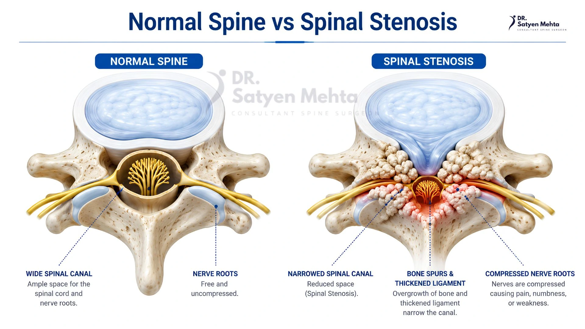
Microscopic discectomy, also known as microdiscectomy, is a surgical procedure that removes the part of a herniated or slipped disc pressing on a spinal nerve.The procedure is commonly performed in the lumbar spine (lower back), where a displaced disc can compress a nerve root and cause symptoms such as sciatica, radiating leg pain, tingling, numbness and muscle weakness.An operating microscope provides magnified visualisation of the surgical area, allowing the surgeon to identify and carefully release the compressed nerve. Only the problematic disc fragment is removed, preserving as much of the remaining disc and surrounding structures as possible.
Dr. Satyen Mehta specialises in microscopic, minimally invasive and endoscopic spine surgery, with more than 25 years of experience dedicated to spinal disorders.
When is Microscopic Discectomy Recommended?
Microscopic discectomy may be considered in patients with:
- Persistent sciatica or radiating leg pain due to lumbar disc herniation.
- Leg pain that does not improve with appropriate medication, physiotherapy or other non-surgical treatment.
- Numbness, tingling or weakness associated with nerve compression.
- Progressive neurological weakness, including foot drop.
- MRI-confirmed lumbar disc herniation that corresponds with clinical symptoms.
- Significant pain or functional limitations affecting everyday activities.
In cases of severe or progressive neurological symptoms, earlier surgical assessment may be necessary. New bladder or bowel dysfunction, or numbness around the groin and saddle area, requires immediate emergency medical evaluation.
How is Microscopic Discectomy Performed?
Microscopic discectomy is generally performed under general anaesthesia. The procedure involves the following steps:
- Anaesthesia and Positioning
The patient is administered general anaesthesia and positioned face down on the operating table to provide access to the lumbar spine.
- Surgical Access
A small incision is made over the affected spinal level. Imaging guidance may be used to confirm the correct location. The operating microscope provides magnified visualisation of the surgical area.
- Nerve Identification and Decompression
The surgeon carefully accesses the affected disc and identifies the compressed nerve root. A small portion of the ligament or bone may be removed if required to reach the disc safely.
- Removal of Herniated Disc Material
The displaced disc fragment pressing against the nerve is carefully removed. The surgeon checks that the nerve has adequate space and is no longer compressed.
- Wound Closure
The surgical instruments are withdrawn, and the incision is closed with appropriate sutures or wound closure material. The patient is shifted to the recovery area for observation.
Benefits of Microscopic Discectomy
When appropriately indicated, microscopic discectomy offers several potential benefits:
- Direct removal of the disc fragment compressing the nerve.
- Magnified visualisation of delicate spinal structures.
- Relief from nerve-related leg pain and sciatica in many patients.
- Preservation of most of the healthy disc.
- Smaller surgical exposure compared with conventional open approaches.
- Potentially earlier mobilisation and recovery.
The expected benefits depend on the patient’s diagnosis, extent of nerve compression and overall health.
Recovery After Microscopic Discectomy
Recovery varies depending on the patient’s condition, the extent of surgery and individual healing.
Immediately After Surgery
Patients are monitored in the recovery area. Mobilisation is generally encouraged once medically appropriate, with assistance from the hospital team.
First 1–2 Weeks
Short walks and gradual movement are encouraged. Some incision-site soreness and fatigue may occur. Heavy lifting and activities involving excessive bending or twisting should be avoided as advised.
Weeks 2–6
Daily activities can gradually increase according to the surgeon’s recommendations. Return to desk-based work may be possible during this period for some patients.
Beyond 6 Weeks
Rehabilitation and strengthening exercises may be introduced or progressed. Return to more demanding physical activities depends on recovery and clinical assessment.
Risks and Possible Complications
Like all surgical procedures, microscopic discectomy carries certain risks. These may include:
- Infection or bleeding.
- Dural tear or leakage of spinal fluid.
- Nerve irritation or injury.
- Persistent pain, numbness or weakness.
- Recurrent disc herniation.
- Blood clots or anaesthesia-related complications.
The surgeon will discuss the potential benefits, risks and alternatives based on the patient’s individual condition before recommending surgery.
Why Consult Dr. Satyen Mehta for Microscopic Spine Surgery?
Dr. Satyen Mehta is an Orthopaedic Spine Surgeon in Mumbai with over 25 years of experience dedicated to treating spinal disorders.
His qualifications and professional background include:
- Gold Medalist in M.S. Orthopaedics.
- Member of the Royal College of Surgeons (MRCS), Glasgow, UK.
- International spine surgery training in South Korea and Australia.
- Fellowship training in spinal deformity corrective surgery and paediatric spine surgery.
- Special interest in microscopic, minimally invasive and endoscopic spine surgery.
His treatment approach focuses on accurate diagnosis, individualised treatment planning and recommending surgery when clinically appropriate.
Frequently Asked Questions
- What is the difference between microscopic discectomy and conventional discectomy?
Microscopic discectomy uses an operating microscope to provide magnified visualisation during surgery. This helps the surgeon identify the nerve and remove the herniated disc material with precision.
- Is microscopic discectomy a major surgery?
It is a specialised spinal surgical procedure involving access to the lumbar disc and nerve root. Although the surgical exposure is relatively small, it requires careful planning, anaesthesia and postoperative monitoring.
- How long does it take to recover from microscopic discectomy?
Many patients gradually resume routine activities over several weeks. Recovery depends on the extent of nerve compression, duration of symptoms, surgical findings and the nature of the patient’s work.
- Does microscopic discectomy permanently cure a slipped disc?
The procedure removes the disc material responsible for the existing nerve compression. However, the remaining disc can develop another herniation, and symptoms may recur in some patients.
- Is microscopic discectomy better than endoscopic discectomy?
Both procedures aim to relieve nerve compression caused by a herniated disc. The appropriate technique depends on the location and nature of the herniation, spinal anatomy and the surgeon’s assessment.
- Is surgery always necessary for a slipped disc?
No. Many patients improve with medication, physiotherapy, activity modification and other non-surgical treatments. Surgery is considered when symptoms persist or neurological concerns make surgical intervention appropriate.